原子の中にある電子は、惑星が太陽の周りを回るように単純な円軌道を動いているわけではありません。現代の量子力学において、電子の居場所は原子軌道(Atomic Orbital)という概念で説明されます。これは電子が特定の領域に存在する確率を数学的な関数(波関数)で表したものであり、原子の化学的性質や結合様式を決定づける極めて重要な要素です。
かつてのモデルでは電子は点のような粒子として捉えられていましたが、実際には波のような性質を持っており、核の周囲に「雲」のように分布しています。この分布の形状やエネルギー状態を理解することが、化学反応や物質の構造を解明する鍵となります。
Key Facts
- 原子軌道とは、電子が存在する確率分布を示す数学的な関数である。
- 1つの軌道には、スピンが異なる最大2つの電子しか入ることができない(パウリの排他原理)。
- 軌道の形状とエネルギーは、3つの量子数(n, ℓ, mℓ)によって定義される。
- 軌道は主にs, p, d, fなどの名称で分類され、それぞれ異なる立体形状を持つ。
- 電子は単一の状態で存在するだけでなく、複数の状態が重なり合った「重ね合わせ」の状態を取ることもある。
原子モデルの変遷:古典的視点から量子力学へ
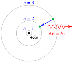
原子構造の理解は、20世紀初頭の試行錯誤から始まりました。1904年に長岡半太郎が提唱したモデルや、1911年にラザフォードが発見した正電荷を持つ中心核の概念を経て、1913年にニールス・ボーアが量子化された角運動量を持つ電子軌道モデルを提示しました。
ボーアのモデルは水素原子のスペクトル線を鮮やかに説明しましたが、多電子原子の挙動や周期表に見られる化学的類似性を完全に説明することはできませんでした。そこで登場したのが、電子を粒子ではなく波として扱う量子力学的なアプローチです。これにより、電子は「軌道(Orbit)」ではなく「軌道(Orbital)」という確率的な分布として定義されるようになりました。
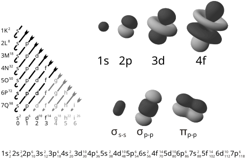
軌道を決定する量子数と種類
電子がどの軌道に属するかは、以下の3つの量子数によって厳密に規定されています。
- 主量子数 (n):電子のエネルギーレベル(電子殻)を示し、1, 2, 3...と正の整数で表されます。
- 方位量子数 (ℓ):軌道の形状(角運動量)を決定します。0からn-1までの値を取り、ℓ=0はs軌道、ℓ=1はp軌道、ℓ=2はd軌道、ℓ=3はf軌道と呼ばれます。
- 磁気量子数 (mℓ):空間的な方向性を決定します。-ℓから+ℓまでの整数値を取ります。
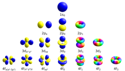
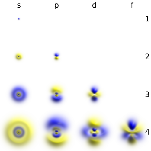
これらの組み合わせにより、電子の分布パターンが決まります。例えば、s軌道は球形であり、p軌道は亜鈴のような形をしています。また、エネルギーレベルが高くなるにつれ、軌道の形状はより複雑になります。
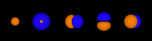
さらに、電子にはスピン量子数 (ms)という固有の性質があり、1つの軌道にはスピンの向きが異なる最大2つの電子が収容されます。これが原子の電子配置の基本ルールとなります。
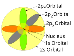
軌道の形状とエネルギーの相関
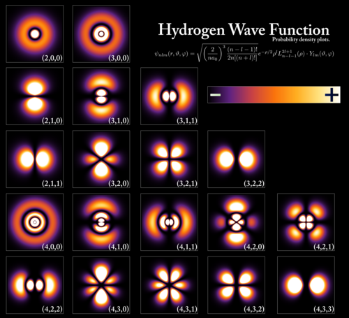
電子の存在確率は、波関数の絶対値の2乗によって計算されます。s軌道は中心対称な球状ですが、nが2以上のs軌道には、電子が存在しない「節(ノード)」と呼ばれる領域が存在します。

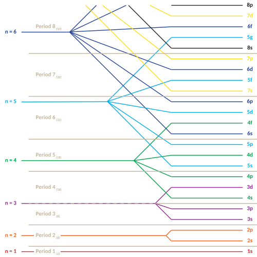
多電子原子の場合、電子同士の反発があるため、エネルギー準位はnだけでなくℓにも依存します。一般的にℓの値が大きいほどエネルギーが高くなります。このため、例えば4s軌道が3d軌道よりも先に満たされるといった、複雑な充填順序(マデ隆則)が生じます。
また、実際の電子は単一の固有状態にあるだけでなく、複数の状態が複素数的な重みで組み合わさった「重ね合わせ状態」として存在することがあります。これにより、数学的な扱いが容易な固有状態の線形結合として、現実的な実数軌道を表現することが可能です。

現代の技術では、走査透過電子顕微鏡(STEM)とエネルギー分散X線分光法(EDX)を用いることで、ストロンチウムなどの原子における1sや2pといった内殻電子の軌道を実験的に可視化することに成功しています。
![Experimentally imaged 1s and 2p core-electron orbitals of Sr, including the effects of atomic thermal vibration and excitation broadening, retrieved from energy dispersive x-ray spectroscopy (EDX) in scanning transmission electron microscopy (STEM).[31]](/images/1c/2d/1c2d5b9ae9a83b8146b9b9840a55c856b0c2ff6d0cb40293d324a06e733be130.jpg)
軌道エネルギーのまとめ
以下に、主要な量子数とそれに対応する軌道の名称および特徴をまとめます。
| 方位量子数 (ℓ) | 軌道記号 | 形状の特徴 | 磁気量子数 (mℓ) の範囲 | 最大収容電子数 (1つの軌道あたり) |
|---|---|---|---|---|
| 0 | s | 球形 | 0 | 2 |
| 1 | p | 亜鈴形(3方向) | -1, 0, 1 | 2 |
| 2 | d | 複雑な四葉形など(5方向) | -2, -1, 0, 1, 2 | 2 |
| 3 | f | さらに複雑な形状(7方向) | -3, -2, -1, 0, 1, 2, 3 | 2 |
特殊な現象:相対論的効果と遷移
非常に原子番号が大きい元素(重元素)では、内殻電子の速度が光速に近づくため、相対論的な効果が無視できなくなります。理論上、原子番号137(フィニマニウムとも呼ばれる)を超えると、非相対論的な量子力学では電子速度が光速を超えてしまう矛盾が生じます。実際には原子核が点電荷ではないため、真空の崩壊や電子・陽電子対の生成が起こる臨界値は原子番号173付近であると考えられています。
また、電子が異なる軌道間を移動することを「遷移」と呼びます。電子が低いエネルギー状態から高い状態へ移るには、そのエネルギー差に正確に一致する光子(エネルギー)を吸収する必要があります。これが、物質固有の吸収スペクトルや放出スペクトルとして観測される理由です。
Frequently Asked Questions
原子軌道と電子軌道(Orbit)は何が違うのですか?
古典的な「軌道(Orbit)」は、惑星のように電子が特定の経路を回っているという概念です。一方、量子力学的な「原子軌道(Orbital)」は、電子がどこに存在するかの「確率分布」を示す領域を指します。電子は特定の線上を動いているのではなく、雲のように空間に分布しています。
なぜs, p, d, fという名前がついているのですか?
これらの名称は、初期の分光分析においてアルカリ金属のスペクトル線が示した特徴から由来しています。sはsharp(鋭い)、pはprincipal(主要な)、dはdiffuse(拡散した)、fはfundamental(基本的な)という英語の頭文字からきています。
1つの軌道に電子が2つまでしか入れないのはなぜですか?
これは「パウリの排他原理」によるものです。量子力学的なルールにより、同じ原子内で4つの量子数(n, ℓ, mℓ, ms)がすべて同一である電子は存在できません。そのため、空間的な軌道(n, ℓ, mℓが同じ)が1つあるとき、スピン(ms)が異なる2つの電子までしか共存できないことになります。
電子が「重ね合わせ」にあるとはどういう意味ですか?
電子が単一の確定した状態(固有状態)ではなく、複数の異なる状態が同時に、ある割合で混ざり合って存在している状態を指します。これは複素数の重みを用いて数学的に記述され、観測されるまで電子の状態は確定していないという量子力学の根本的な性質を表しています。
相対論的効果は化学にどのような影響を与えますか?
非常に重い元素では、相対論的効果によってs軌道などのエネルギー準位が低下し、軌道が収縮します。これにより、金(Au)が特有の黄色い光沢を持つことや、水銀(Hg)が室温で液体であることなど、周期表の下方に位置する元素の特異な化学的性質に影響を与えています。
References
- Orchin, Milton; MacOmber, Roger S.; Pinhas, Allan R.; Wilson, R. Marshall (2005). "Atomic Orbital Theory". The Vocabulary and Concepts of Organic Chemistry. pp. 1–24. :10.1002/0471713740.ch1. .
- Daintith, J. (2004). Oxford Dictionary of Chemistry. New York: Oxford University Press. pp. 407–409. .
- Griffiths, David (1995). Introduction to Quantum Mechanics. Prentice Hall. pp. 190–191. .
- Levine, Ira (2000). Quantum Chemistry (5 ed.). Prentice Hall. pp. 144–145. .
- Laidler, Keith J.; Meiser, John H. (1982). Physical Chemistry. Benjamin/Cummings. p. 488. .
- Atkins, Peter; de Paula, Julio; Friedman, Ronald (2009). Quanta, Matter, and Change: A Molecular Approach to Physical Chemistry. Oxford University Press. p. 106. .
- Feynman, Richard; Leighton, Robert B.; Sands, Matthew (2006). The Feynman Lectures on Physics – The Definitive Edition. Vol. 1, lecture 6. Pearson PLC, Addison Wesley. p. 11. .
- , .
- Levine, Ira N. (1991). Quantum Chemistry (4th ed.). Prentice-Hall. p. 262. .
Therefore, the wave function of a system of identical interacting particles must not distinguish among the particles.
- Mulliken, Robert S. (July 1932). "Electronic Structures of Polyatomic Molecules and Valence. II. General Considerations". Physical Review. 41 (1): 49–71. :1932PhRv...41...49M. :10.1103/PhysRev.41.49.
📸 フォトギャラリー