An ECG in a person with a single-chamber pacemaker to the atrium. Note the circle around one of the sharp electrical spikes in the position where the P wave would be expected.
← ホームへ戻る

ペースメーカーの仕組みと進化心拍リズムを制御する医療技術の全貌

🗓 2026年8月16日

心臓が適切なリズムで拍動しなくなったとき、生命を維持するために不可欠なのがペースメーカーです。これは、電気刺激を用いて心筋を収縮させ、心拍数を適切に維持するデバイスです。初期の巨大な外部装置から、現代の心臓内に直接留置する超小型デバイスに至るまで、この技術は劇的な進化を遂げてきました。

Key Facts

  • 基本機能:心臓の電気信号を感知し、必要に応じて電気刺激を与えて心拍を維持する。
  • 多様な種類:単腔、二腔、そして心不全治療に用いられるbiventricular(両心室)ペースメーカーなどが存在する。
  • 最新トレンド:リード(電極線)を必要としない「リードレスペースメーカー」が登場し、低侵襲化が進んでいる。
  • 多機能化:除細動機能を持つICD(植込み型除細動器)や、心不全を改善するCRT(心臓再同期療法)へと発展している。

ペースメーカーの基本原理と動作モード

ペースメーカーは、心臓の電気的な活動を常に監視(センシング)し、心拍が設定値を下回った場合にのみ電気パルスを送信します。この動作は、どの心腔(心房や心室)を刺激し、どこから信号を感知するかによって定義されます。

例えば、心房のみを刺激する単腔ペースメーカーの場合、心電図では本来のP波があるべき位置に鋭い電気的スパイクが確認されます。
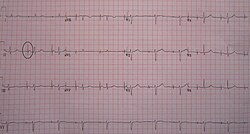
An ECG in a person with a single-chamber pacemaker to the atrium. Note the circle around one of the sharp electrical spikes in the position where the P wave would be expected.

より高度な二腔ペースメーカーでは、心房と心室の両方を制御し、より自然な心拍リズムを再現することが可能です。
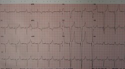
An ECG of a person with a dual-chamber pacemaker

刺激の閾値と依存性

心筋を収縮させるために必要な最小限の電気量を「閾値」と呼びます。もし患者自身の心拍が完全に停止しており、ペースメーカーの刺激がなければ心停止に至る状態を「ペースメーカー依存」と呼びます。これは、刺激強度を下げた際に心室からの自発的なリズム(逃避リズム)が現れないことで判断されます。
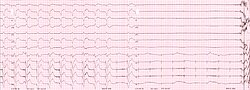
ECG rhythm strip of a threshold determination in a patient with a temporary (epicardial) ventricular pacemaker. The epicardial pacemaker leads were placed after the patient collapsed during aortic valve surgery. In the first half of the tracing, pacemaker stimuli at 60 beats per minute result in a wide QRS complex with a right bundle branch block pattern. Progressively weaker pacing stimuli are administered, which results in asystole in the second half of the tracing. At the end of the tracing, distortion results from muscle contractions due to a (short) hypoxic seizure. Because decreased pacemaker stimuli do not result in a ventricular escape rhythm, the patient can be said to be pacemaker-dependent and needs a definitive pacemaker.

デバイスの構造と植込み手法

従来のペースメーカーは、本体(ジェネレーター)を皮下に植え込み、そこから「リード」と呼ばれる電極線を静脈経由で心臓内部へ導く構造になっています。
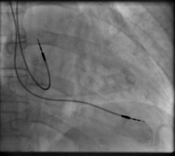
Right atrial and right ventricular leads as visualized under x-ray during a pacemaker implant procedure. The atrial lead is the curved one making a U shape in the upper left part of the figure.

植込み後の状態を確認するためには、胸部X線写真が用いられ、リードが右心房や右心室に正しく配置されているかを確認します。
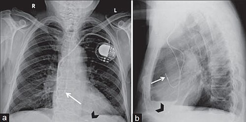
Posteroanterior and lateral chest radiographs of a pacemaker with normally located leads in the right atrium (white arrow) and right ventricle (black arrowhead), respectively

高度な治療デバイス:CRTとICD

心不全などの重症例では、右心房、右心室に加えて、冠静脈洞を通じて左心室にもリードを配置する「心臓再同期療法(CRT)」が行われます。また、致死的な頻脈(心室細動など)が発生した際に電気ショックで停止させる機能を持つのがICD(植込み型除細動器)です。これらのデバイスは、通常のペースメーカーよりもジェネレーターのサイズが大きくなる傾向があります。
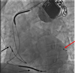
Three leads can be seen in this example of a cardiac resynchronization device: a right atrial lead (solid black arrow), a right ventricular lead (dashed black arrow), and a coronary sinus lead (red arrow). The coronary sinus lead wraps around the outside of the left ventricle, enabling pacing of the left ventricle. Note that the right ventricular lead in this case has two thickened aspects that represent conduction coils and that the generator is larger than typical pacemaker generators, demonstrating that this device is both a pacemaker and a cardioverter-defibrillator, capable of delivering electrical shocks for dangerously fast abnormal ventricular rhythms.

リードレスペースメーカーの登場

近年では、リードを一切使用せず、カプセル状の小型デバイスをカテーテルで直接心室内に留置する「リードレスペースメーカー」が実用化されました。これにより、リード断線や感染症のリスクを低減し、手術の侵襲性を大幅に下げることが可能となりました。
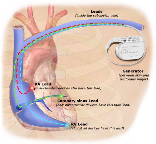
Illustration of implanted cardiac pacemaker showing locations of cardiac pacemaker leads

ペースメーカーの歴史的変遷

心臓への電気刺激の試みは19世紀末にまで遡ります。1889年にジョン・アレクサンダー・マクウィリアムが心停止状態の心臓に電気刺激を与え、心室収縮を誘発したことが先駆けとなりました。

その後、1920年代にはポータブル装置が開発され、1958年にはスウェーデンのルネ・エルムクヴィストとオーケ・セニングによって世界初の完全植込み型ペースメーカーが誕生しました。最初の患者であるアルネ・ラーソン氏は、生涯にわたって26台ものデバイスを交換しながら、86歳まで生存しました。
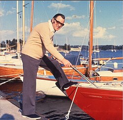
In 1958, Arne Larsson (1915–2001) became the first person to receive an implantable pacemaker. He had 26 devices during his life and campaigned for other patients needing pacemakers.

初期の外部装置は非常に大きく、1961年当時のモデルなどは現在の基準からは想像できないサイズでした。
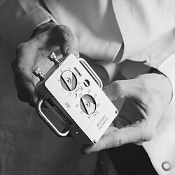
An external Medtronic pacemaker in 1961

電源技術の革新

初期のデバイスは寿命が短く、頻繁な交換手術が必要でした。1970年代には一時的にプルトニウムを用いた原子力電池が導入されましたが、その後、1974年にアンソニー・アドゥッチらによって開発されたリチウム・ヨウ素電池が標準となりました。これにより、デバイスの寿命は飛躍的に延び、現代の長期的な使用が可能となりました。

The first lithium-iodide cell-powered pacemaker. Invented by Anthony Adducci and Art Schwalm. Cardiac Pacemakers Inc. 1972[89]

管理とライフスタイルへの影響

植込み後の患者は、定期的なチェックアップが必要です。現代では、クリニックに足を運ばなくてもデバイスの状態を監視できるリモートモニタリングシステムが普及しています。
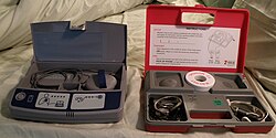
Two types of remote monitoring devices used by pacemaker patients

また、強い磁場やMRI検査、一部の電化製品(IHクッキングヒーターなど)による電磁干渉に注意が必要です。ただし、最近ではMRI検査に対応した「MRI条件付き」デバイスも開発されています。

動物への応用

ペースメーカー技術は人間だけでなく、犬、猫、馬などの動物医療にも応用されています。特に、人間用の寄贈デバイスを再利用することで、飼い主の費用負担を軽減する取り組みも行われています。動物の場合、術後の安静維持が困難であるため、特に注意深い管理が求められます。

まとめ:ペースメーカーの特性一覧

ペースメーカーの種類と特徴
種類 主な目的 特徴
単腔ペースメーカー 徐脈の改善 心房または心室のどちらか一方のみを刺激
二腔ペースメーカー 生理的な心拍維持 心房と心室の両方を協調して刺激
CRT (心臓再同期療法) 心不全の改善 左心室を含む3箇所を刺激し、収縮を同期させる
ICD (植込み型除細動器) 突然死の防止 ペースメーカー機能に加え、致死的不整脈への電気ショック機能を持つ
リードレスペースメーカー 低侵襲な徐脈治療 リードがなく、心臓内に直接留置する超小型設計

Frequently Asked Questions

ペースメーカーを植え込むと生活に制限はありますか?

多くの日常生活は可能ですが、強い磁場を発生させる機器やMRI検査、一部の工業用機器などの使用に制限がある場合があります。最新のデバイスではMRI対応のものも増えていますが、必ず医師に確認してください。

電池が切れたらどうなるのでしょうか?

電池が完全に切れる前に、定期検診で残量がチェックされます。寿命が近づいた段階で、ジェネレーター(本体)のみを交換する手術が行われるため、突然停止するリスクは極めて低くなっています。

リードレスペースメーカーのメリットは何ですか?

リード(電極線)を使用しないため、リードの断線や脱落、リード沿いの感染症といった合併症のリスクを回避できます。また、皮膚の下に本体を植え込む「ポケット」が不要なため、外見上の違和感や術後の痛みが少ないのが特徴です。

ICDと通常のペースメーカーの違いは何ですか?

通常のペースメーカーは「心拍が遅すぎる(徐脈)」ことを防ぐためのものです。一方、ICDは徐脈への対応に加え、「心拍が速すぎる(致死的な頻脈)」ことを検知し、電気ショックを与えて正常なリズムに戻す機能を持っています。

動物にもペースメーカーは使えるのですか?

はい、犬や猫、馬などの動物にも使用されています。人間と同様に心不全や徐脈の治療に有効であり、一部では人間用の寄贈デバイスを再利用してコストを抑える運用も行われています。

References

  1. "Devices@FDA". Accessdata.fda.gov. Retrieved 2022-04-27.
  2. Product Performance Report – Cardiac Rhythm Management (PDF) (Report). St. Jude Medical. October 2008. Archived from the original (PDF) on 2022-08-19. Retrieved 2022-04-27.
  3. "UpToDate". Uptodate.com. Retrieved 2022-08-02.
  4. "Pacemakers". Retrieved 28 July 2022.
  5. "Overview of Pacemakers and Implantable Cardioverter Defibrillators (ICDs) – Health Encyclopedia – University of Rochester Medical Center". Urmc.rochester.edu. Retrieved 2022-08-02.
  6. "Biventricular Pacemaker". Cleveland Clinic. Retrieved 2022-08-02.
  7. Eich C, Bleckmann A, Paul T (October 2005). "Percussion pacing in a three-year-old girl with complete heart block during cardiac catheterization". Br J Anaesth. 95 (4): 465–67. :10.1093/bja/aei209.  16051649.
  8. Shah, Maully; Rhodes, Larry; Kaltman, Jonathan (2017). Cardiac Pacing and Defibrillation in Pediatric and Congenital Heart Disease. John Wiley & Sons.  .
  9. Valchanov, Kamen; Jones, Nicola; Hogue, Charles W. (2018). Core Topics in Cardiothoracic Critical Care. Cambridge University Press.  .[]
  10. "Pacemakers, Patient and Public Information Center: Heart Rhythm Society". Archived from the original on 2010-06-19.

📸 フォトギャラリー

An ECG in a person with a single-chamber pacemaker to the atrium. Note the circle around one of the sharp electrical spikes in the position where the P wave would be expected.
An ECG of a person with a dual-chamber pacemaker
ECG rhythm strip of a threshold determination in a patient with a temporary (epicardial) ventricular pacemaker. The epicardial pacemaker leads were placed after the patient collapsed during aortic valve surgery. In the first half of the tracing, pacemaker stimuli at 60 beats per minute result in a wide QRS complex with a right bundle branch block pattern. Progressively weaker pacing stimuli are administered, which results in asystole in the second half of the tracing. At the end of the tracing, distortion results from muscle contractions due to a (short) hypoxic seizure. Because decreased pacemaker stimuli do not result in a ventricular escape rhythm, the patient can be said to be pacemaker-dependent and needs a definitive pacemaker.
Right atrial and right ventricular leads as visualized under x-ray during a pacemaker implant procedure. The atrial lead is the curved one making a U shape in the upper left part of the figure.
Three leads can be seen in this example of a cardiac resynchronization device: a right atrial lead (solid black arrow), a right ventricular lead (dashed black arrow), and a coronary sinus lead (red arrow). The coronary sinus lead wraps around the outside of the left ventricle, enabling pacing of the left ventricle. Note that the right ventricular lead in this case has two thickened aspects that represent conduction coils and that the generator is larger than typical pacemaker generators, demonstrating that this device is both a pacemaker and a cardioverter-defibrillator, capable of delivering electrical shocks for dangerously fast abnormal ventricular rhythms.
Posteroanterior and lateral chest radiographs of a pacemaker with normally located leads in the right atrium (white arrow) and right ventricle (black arrowhead), respectively
Two types of remote monitoring devices used by pacemaker patients
In 1958, Arne Larsson (1915–2001) became the first person to receive an implantable pacemaker. He had 26 devices during his life and campaigned for other patients needing pacemakers.
An external Medtronic pacemaker in 1961
Illustration of implanted cardiac pacemaker showing locations of cardiac pacemaker leads
The first lithium-iodide cell-powered pacemaker. Invented by Anthony Adducci and Art Schwalm. Cardiac Pacemakers Inc. 1972[89]