地球上のほぼすべての生命体において、設計図の役割を果たすのがDNA(デオキシリボ核酸)です。この複雑な分子は、生物の成長、発達、そして次世代への形質の継承に必要なあらゆる情報を保持しています。DNAは単なる化学物質ではなく、精密な情報ストレージとして機能し、細胞内の指令塔として生命活動を制御しています。
Key Facts
- 基本構造: 2本のポリヌクレオチド鎖が互いに巻き付いた二重らせん構造を持つ。
- 構成要素: リン酸、糖(デオキシリボース)、および4種類の塩基(A, T, G, C)からなるヌクレオチドが基本単位。
- 情報の保存: 塩基の配列順序が遺伝コードとなり、タンパク質の合成指示書として機能する。
- パッケージング: 長大なDNA鎖はヒストンなどのタンパク質に巻き付き、凝縮されて染色体を形成する。
- 複製能力: 自己複製により、細胞分裂時に正確に遺伝情報をコピーして娘細胞へ受け継ぐ。
DNAの化学的構造と形態
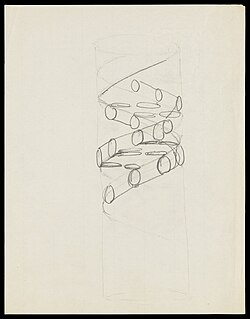
DNAの最も象徴的な形態は、2本の鎖が螺旋状に絡み合った二重らせん構造です。この構造により、膨大な情報をコンパクトに収納しつつ、安定して保持することが可能になります。
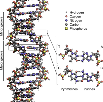
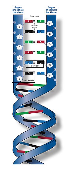
DNAの基本単位はヌクレオチドであり、これは糖、リン酸、そして窒素含有塩基の3つが結合したものです。塩基にはアデニン(A)、チミン(T)、グアニン(G)、シトシン(C)の4種類があり、これらが特定のペア(AとT、GとC)で水素結合を形成することで、2本の鎖が結びついています。
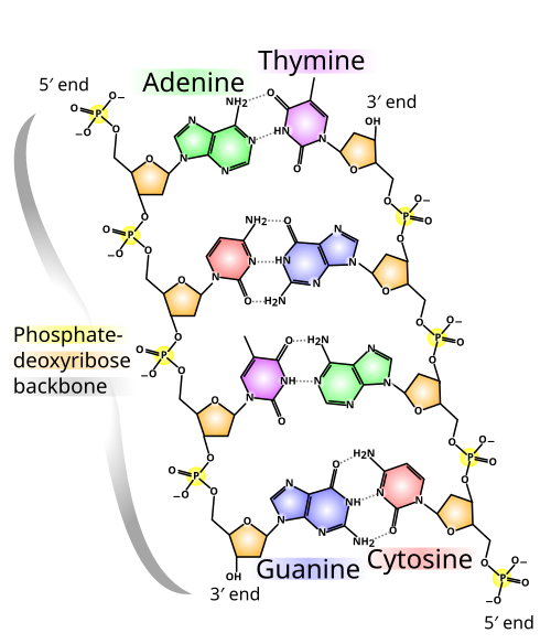
化学的な視点で見ると、DNAの末端には5'(5プライム)側と3'(3プライム)側という方向性があります。5'末端にはリン酸基が、3'末端にはヒドロキシ基(-OH)が露出しており、この方向性が複製や転写のプロセスにおいて極めて重要になります。
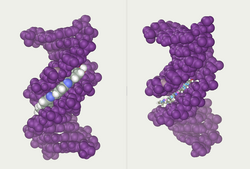
らせん構造の間には、幅の広い「大溝(メジャーグルーブ)」と幅の狭い「小溝(マイナーグルーブ)」が存在します。これらの溝は、特定のタンパク質がDNA配列を認識して結合するための重要なアクセスポイントとなります。
![A section of DNA. The bases lie horizontally between the two spiraling strands[15] (animated version).](/images/34/c8/34c83b76b74f5b8c3899f7dac5b3acf147034c91a9053d48307e6e188bb01dd8.gif)
多様なDNA構造
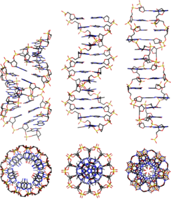
一般的に知られるB-DNA以外にも、環境や条件によってA-DNAやZ-DNAといった異なる形態をとることがあります。また、テロメアなどの特定の領域では、4本の鎖が組み合わさった「G-クアドルプレックス」という特殊な構造を形成することもあります。
![DNA quadruplex formed by telomere repeats. The looped conformation of the DNA backbone is very different from the typical DNA helix. The green spheres in the center represent potassium ions.[62]](/images/f9/7b/f97bdad397d975d526b26e83e8268975877e680d4de5abb13d2cd78e9923ee40.png)
ゲノムの組織化とパッケージング
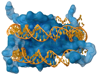
ヒトの細胞一つに含まれるDNAは非常に長く、そのままでは細胞核に収まりません。そのため、DNAはヒストンと呼ばれる塩基性タンパク質に巻き付き、高度に凝縮された構造をとります。この凝縮体こそが染色体であり、細胞分裂時に遺伝情報を効率よく分配するための形態です。
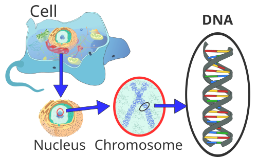
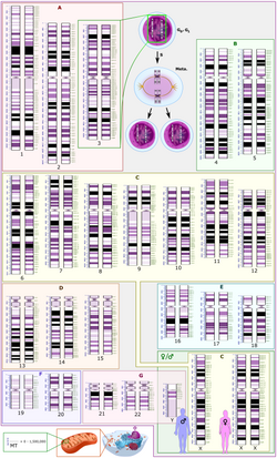
染色体は、常染色体と性染色体に分けられ、それぞれが特定の遺伝子群を保持しています。ミトコンドリアなどの細胞小器官にも、独自の環状DNA(ミトコンドリアゲノム)が存在しています。
生命維持を支えるDNAの動的プロセス
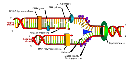
DNAは静止した設計図ではなく、常に動的に利用されています。最も重要なプロセスの一つが複製です。ヘリカーゼなどの酵素が二重らせんを解き、DNAポリメラーゼが元の鎖を鋳型にして新しい相補的な鎖を合成します。この際、リーディング鎖は連続的に、ラギング鎖は岡崎フラグメントと呼ばれる断片的に合成されます。
また、DNAに書き込まれた情報は転写というプロセスを経てRNAにコピーされ、さらにそれが翻訳されることで、生命の実体であるタンパク質が合成されます。この一連の流れは「セントラルドグマ」として知られています。
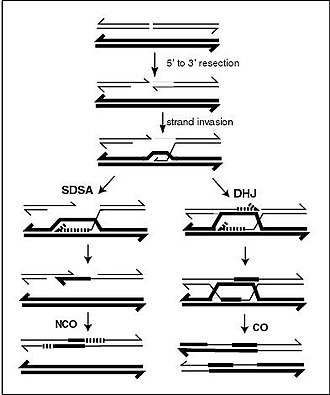
さらに、減数分裂などの過程では、相同染色体間でDNAの一部が入れ替わる遺伝的組換えが起こります。これにより遺伝的多様性が生まれ、生物の進化を促進しています。
![The lambda repressor helix-turn-helix transcription factor bound to its DNA target[117]](/images/4f/19/4f19dd6e4b5f8b0f8d309dc0c531fefe9f9e0682ac8febe372ba13a808af1d61.png)
![The restriction enzyme EcoRV (green) in a complex with its substrate DNA[121]](/images/a3/29/a32951a2d1b0e59be43e9b2f0451d0596c8a558fd72620d395f745ccd8c76a02.png)
DNAの損傷と技術的応用
DNAは化学的に安定していますが、紫外線や化学物質、代謝過程で生じる活性酸素などによって損傷を受けることがあります。例えば、タバコの煙に含まれるベンゾ[a]ピレンなどの発がん物質は、DNAに共有結合して構造を歪ませ、突然変異を引き起こす原因となります。
![A covalent adduct between a metabolically activated form of benzo[a]pyrene, the major mutagen in tobacco smoke, and DNA[82]](/images/75/05/7505cde20a195b97f8e281742fd68f0453b8847d5920e4948d68af1727e28877.png)
現代科学では、DNAの特性を利用した多様な技術が開発されています。個人の塩基配列の違いを利用したDNAプロファイリング(鑑定)や、特定の配列を設計してナノスケールの構造体を作るDNAナノテクノロジーなどがその例です。
![The DNA structure at left (schematic shown) will self-assemble into the structure visualized by atomic force microscopy at right. DNA nanotechnology is the field that seeks to design nanoscale structures using the molecular recognition properties of DNA molecules.[171]](/images/59/0e/590e8e3a3c3df8b68bc86531bf7712fb5eee8d353d115279b253174ce1609d68.png)
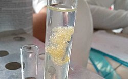
また、純粋なDNAを抽出すると、白く粘り気のある塊として観察することができます。これはDNAが長いポリマーであるためです。
DNA発見の歴史
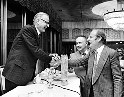
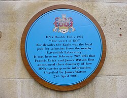
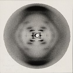
DNAの構造解明は、20世紀半ばの科学的競争の産物でした。ロザリンド・フランクリンとレイモンド・ゴスリングによるX線回折写真(フォト51)が決定的な証拠となり、それを基にジェームズ・ワトソンとフランシス・クリックが1953年に二重らせんモデルを提唱しました。
DNAの基本特性まとめ
| 項目 | 詳細 | 役割 |
|---|---|---|
| ヌクレオチド | 糖・リン酸・塩基の複合体 | DNAの基本構成単位 |
| 塩基対 (A-T, G-C) | 相補的な水素結合 | 情報の正確なコピーと保持 |
| 二重らせん | 右巻きの螺旋構造 | 情報の高密度保存と安定化 |
| 染色体 | DNAとヒストンの凝縮体 | 細胞内での効率的な配置と分配 |
Frequently Asked Questions
DNAとRNAの決定的な違いは何ですか?
主な違いは3点あります。第一に糖の種類で、DNAはデオキシリボース、RNAはリボースを使用します。第二に塩基で、DNAはチミン(T)を持ちますが、RNAではウラシル(U)に置き換わっています。第三に構造で、DNAは通常二重鎖ですが、RNAは主に一本鎖として存在します。
突然変異はどのようにして起こるのですか?
DNAの複製ミスや、外部からの放射線、化学物質による塩基の損傷によって起こります。通常は修復酵素がこれらを修正しますが、修正しきれなかった変更が固定されると突然変異となり、それが形質変化や疾患の原因となることがあります。
DNAナノテクノロジーとはどのような技術ですか?
DNAの相補的な塩基対形成(AはTと、GはCと結合する)という性質を利用して、あらかじめ設計した配列のDNA鎖を自己組織化させ、ナノメートルサイズの箱や幾何学的な構造物を構築する技術です。
ゲノムとは具体的に何を指しますか?
ある生物が持つすべての遺伝情報のセットのことです。ヒトの場合、核にある22対の常染色体と1対の性染色体、およびミトコンドリアDNAのすべてを合わせたものがゲノムに相当します。
References
- "deoxyribonucleic acid". . Merriam-Webster. 1032680871.:
- Alberts B, Johnson A, Lewis J, Raff M, Roberts K, Walter P (2014). Molecular Biology of the Cell (6th ed.). Garland. p. Chapter 4: DNA, Chromosomes and Genomes. . Archived from the original on 14 July 2014.
- Purcell A. "DNA". Basic Biology. Archived from the original on 5 January 2017.
- "Uracil". Genome.gov. Archived from the original on 19 October 2019. Retrieved 21 November 2019.
- Russell P (2001). iGenetics. New York: Benjamin Cummings. .
- Saenger W (1984). Principles of Nucleic Acid Structure. New York: Springer-Verlag. .
- Alberts B, Johnson A, Lewis J, Raff M, Roberts K, Peter W (2002). Molecular Biology of the Cell (Fourth ed.). New York and London: Garland Science. . 145080076. Archived from the original on 1 November 2016.
- , (April 1953). "Molecular structure of nucleic acids; a structure for deoxyribose nucleic acid" (PDF). Nature. 171 (4356): 737–38. :1953Natur.171..737W. :10.1038/171737a0. 0028-0836. 13054692. 4253007. Archived (PDF) from the original on 4 February 2007.
- Berg J, Tymoczko J, Stryer L (2002). Biochemistry. W.H. Freeman and Company. .
- IUPAC-IUB Commission on Biochemical Nomenclature (CBN) (December 1970). "Abbreviations and Symbols for Nucleic Acids, Polynucleotides and their Constituents. Recommendations 1970". The Biochemical Journal. 120 (3): 449–54. :10.1042/bj1200449. 0306-3283. 1179624. 5499957. Archived from the original on 5 February 2007.
📸 フォトギャラリー